Content
The fleeting amaurosis also known as temporary or transient visual loss, is the loss, darkening or blurring of vision that can last from seconds to minutes, and can be only in one or both eyes. The reason this happens is the lack of oxygen-rich blood for the head and eyes.
However, fleeting amaurosis is only a symptom of other conditions, which are usually stress and migraine attacks, for example, but which can also be associated with serious conditions such as atherosclerosis, thromboemboli and even a stroke (stroke) .
In this way, treatment for fleeting amaurosis is done by eliminating what the cause is, and for that reason, it is important to seek medical attention as soon as the problem is perceived, so that the appropriate treatment is started and the chances of sequelae by lack of oxygenation in the tissues.
Possible causes
The main cause of fleeting amaurosis is the lack of oxygen-rich blood in the eye region, made by the artery called the carotid artery, which in this case cannot carry the required amount of oxygenated blood.
Typically, fleeting amaurosis occurs due to the presence of the following conditions:
- Migraine attacks;
- Stress;
- Panic attack;
- Vitreous hemorrhage;
- Hypertensive crisis;
- Anterior ischemic optic neuropathy;
- Convulsions;
- Vertebrobasilar ischemia;
- Vasculitis;
- Arteritis;
- Atherosclerosis;
- Hypoglycemia;
- Vitamin B12 deficiency;
- Smoking;
- Thiamine deficiency;
- Corneal trauma;
- Cocaine abuse;
- Infections with toxoplasmosis or cytomegalovirus;
- High plasma viscosity.
The fleeting amaurosis is always temporary, and therefore the vision returns to normal in a few minutes, in addition to not usually leaving any sequelae, however it is necessary that a doctor is sought even if the amaurosis has lasted a few seconds, so that what can be investigated caused it.
In rare cases, the person may experience symptoms before the fleeting amaurosis occurs, but when it does, mild pain and itchy eyes are reported.
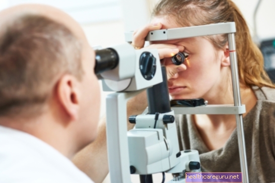
How to confirm the diagnosis
The diagnosis of fleeting amaurosis is made by the general practitioner or ophthalmologist through the patient's report, a physical examination that will check if there is any injury caused by falls or blows, followed by an ophthalmological examination in order to observe possible eye injuries.
Tests such as complete blood count, C-reactive protein (CRP), lipid panel, blood glucose level, echocardiogram and an evaluation of the carotid vein circulation may also be necessary, which can be done by doppler or angioresonance, so that the that caused amaurosis and thus start the appropriate treatment.
How the treatment is done
Treatment for fleeting amaurosis aims to eliminate its cause, and this is usually done with the use of drugs such as antiplatelet agents, antihypertensives and corticosteroids, in addition to dietary reeducation and, if necessary, exercises to eliminate excess weight and start the practice. relaxation techniques.
However, in more severe cases where the carotid artery is seriously obstructed, whether due to stenosis, atherosclerosis or clots, surgery for carotid endarterectomy or angioplasty may be indicated to reduce the risks of a possible stroke. See how angioplasty is done and what the risks are.
Created by: Tua Saúde Editorial Team
Bibliography>
- ANN NEUROL. Amaurosis fugx and ocular infarction in adolescents and young adults. 1989. Available at:. Accessed on 21 Sep 2020
- STROKE. Spectrum of transient visual symptoms in a transient ischemic attack cohort. 2013. Available at:. Accessed on 21 Sep 2020
- J NEUROL NEUROSURG PSYCHIATRY. Clinical features of transient monocular blindness and the likelihood of atherosclerotic lesions of the internal carotid artery. 2001. Available at:. Accessed on 21 Sep 2020
- N ENGL J MED. Clinical and angiographic features of carotid transient ischemic attacks. 1977. Available at:. Accessed on 21 Sep 2020
- STATPEARLS. Amaurosis Fugax. 2020. Available at:. Accessed on 21 Sep 2020
- CLIN OPHTHALMOL. Amaurosis fugx: risk factors and prevalence of significant carotid stenosis. 2016. Available at:. Accessed on 21 Sep 2020


.jpg)