Content
Cardiac catheterization is a procedure that can be used to diagnose or treat heart disease, which involves the introduction of a catheter, which is an extremely thin flexible tube, in the artery of the arm, or leg, up to the heart. Cardiac catheterization may also be known as coronary angiography.
This type of procedure can be indicated both for the diagnosis of some heart problems, as well as for the treatment of infarction or angina, as it examines the interior of blood vessels and the heart, being able to detect and remove accumulations of fatty plaques or lesions in these regions.

How cardiac catheterization is done
What is it for
Cardiac catheterization serves to diagnose and / or treat various cardiac conditions, among which we can highlight:
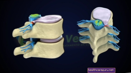
- Assess whether the coronary arteries, which supply the heart muscle, are clogged or not;
- Clear arteries and valves due to the accumulation of fatty plaques;
- Check for damage to the valves and heart muscle;
- Check for changes in the anatomy of the heart not confirmed by other tests;
- Show in detail, if any, a congenital malformation in newborns and children.
Cardiac catheterization can be performed combined with other techniques such as coronary angioplasty, a technique used to unblock the coronary vessel and can be performed with a stent implant (metallic prosthesis) or only with the use of a balloon, which with high pressures, pushes the plates, opening the vase. Learn more about how angioplasty is performed.
It can also be done in conjunction with percutaneous balloon valvuloplasty, used to treat diseases such as heart valves such as pulmonary stenosis, aortic stenosis and mitral stenosis. Also, learn more details about indications of how valvuloplasty is performed.
How cardiac catheterization is done
Cardiac catheterization is done by inserting a catheter or tube into the heart. The step by step is:
- Local anesthesia;
- Making a small opening for the catheter to enter the skin of the groin or forearm at the wrist or elbow;
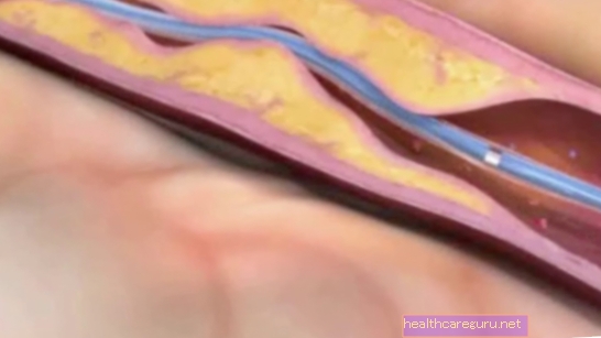
- Insertion of the catheter in the artery (usually, radial, femoral or bracheal) that will be guided by the specialist doctor, to the heart;
- Location of the entrances of the right and left coronary arteries;
- Injection of an iodine-based substance (contrast) that allows the visualization of the arteries and their points of obstruction by X-rays;
- Contrast injection into the left ventricle, allowing visualization of cardiac pumping.
The exam does not cause pain. The most that can happen is that the patient feels some discomfort in the bite of anesthesia and a passing wave of heat in the chest when injecting the contrast.
The duration of the examination varies according to how easy it is to catheterize the target, being generally longer in patients who have already undergone myocardial revascularization surgery. Normally, the exam does not take more than 30 minutes, and it is necessary to remain at rest for a few hours and, if there is no problem, you can go home if you have only performed the catheterization without another associated procedure.
What care is needed
Generally, for a scheduled catheterization, it is necessary to fast for 4 hours before the exam, and try to rest. In addition, only medicines prescribed by the cardiologist should be kept in use, avoiding remedies that were not prescribed, including home remedies and teas. Check out what are the main cares that should be taken before and after surgery.
Generally, recovery from the procedure is quick, and when there are no other complications that prevent it, the patient is discharged from the hospital the next day with a recommendation to avoid vigorous exercise or to lift weights over 10 kg in the first 2 weeks after the procedure.
Possible risks of catheterization
Despite being very important and generally safe, this procedure can bring some health risks, such as:
- Bleeding and infection at the catheter insertion site;
- Blood vessel damage;
- Allergic reaction to the contrast used;
- Irregular heartbeat or arrhythmia, which may go away on its own, but may need treatment in case of persistence;
- Blood clots that can trigger stroke or heart attack;
- Drop in blood pressure;
- Accumulation of blood in the sac that surrounds the heart, which can prevent the heart from beating normally.
The risks are minimal when the exam is scheduled, moreover, it is usually done in reference hospitals in cardiology and well equipped, containing cardiologists and cardiac surgeons, by sus or private.
These risks can happen, especially, in diabetics, patients with kidney diseases and individuals over 75 years old, or in those more severe and acute patients with myocardial infarction.