Content
In most cases, the blue nevus is a benign skin change that is not life-threatening and therefore does not need to be removed. However, there are some cases where the development of malignant cells appears at the site, but this is only more common when the blue nevus is very large or increases in size rapidly.
The blue nevus is similar to a wart and develops due to the accumulation, in the same place, of several melanocytes, which are the skin cells responsible for the darkest color. As these cells are present in a deeper layer of the skin, their color does not appear completely and, therefore, they appear to have a blue color, which can vary even dark gray.
This type of alteration in the skin is more frequent on the head, neck, bottom of the back, hands or feet, being easily evaluated by the dermatologist, and can appear in people of all ages, being more frequent in children and young adults.
How blue nevus is diagnosed
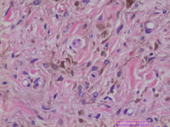
The diagnosis of blue nevus is easy, being performed by the dermatologist only after observing the characteristics presented by the nevus, such as small size, between 1 and 5 mm, rounded shape and raised or smooth surface. In case of changes in the nevus, it may be necessary to perform a differential diagnosis by means of biopsy, in which the cellular characteristics of the nevus are observed.
The differential diagnosis of the blue nevus is made for melanoma, dermatofibroma, plantar wart and tattoo.
When to go to the doctor
Although the blue nevus is almost always a benign alteration, it is important to be aware of its characteristics, especially when it appears after the age of 30. Therefore, it is recommended to go to the doctor when:
- The nevus rapidly increases in size;
- Development for shape with irregular edges;
- Changes in color or appearance of various colors;
- Asymmetric stain;
- The nevus starts to itch, hurt or bleed.
Thus, whenever the nevus changes after diagnosis, it is advisable to consult the dermatologist again for further exams and, if necessary, perform a minor surgery to remove the nevus. This surgery can be done in the dermatologist's office under local anesthesia, and it is not necessary to do any type of preparation. Usually, the blue nevus is removed in about 20 minutes and then sent to the laboratory to assess the presence of malignant cells.
When malignant cells are found after removing the blue nevus, the doctor assesses its degree of development and, if it is high, may recommend repeating the surgery to remove some of the tissue that was around the nevus, to remove all cancer cells. Learn to identify the signs and symptoms indicative of skin cancer.
Created by: Tua Saúde Editorial Team
Bibliography>
- AMERICAN OSTEOPATHIC COLLEGE OF DERMATOLOGY (AOCD). Blue Nevus. Available in: . Accessed on Dec 12, 2019
- ZEMBOWICZ, Artur; PHADE, Pushkar A. Blue Nevi and Variants: An Update. Arch Pathol Lab Med. Vol 135. 2011


.jpg)