Content

The septate uterus is a congenital uterine malformation in which the uterus is divided in two due to the presence of a membrane, also called a septum. The presence of this septum does not lead to the appearance of signs or symptoms, however it can be identified during routine exams.
Although it does not cause symptoms, the septate uterus can make pregnancy difficult and, therefore, it is important that it be identified and treated according to the gynecologist's guidance, and a surgical procedure may be indicated to remove the wall that separates the uterus. .
How to identify
The septate uterus in most cases does not lead to the appearance of signs or symptoms, being only identified through routine gynecological exams. In addition, when the woman has difficulty getting pregnant or has several spontaneous abortions, it is possible that it is indicative of uterine changes.
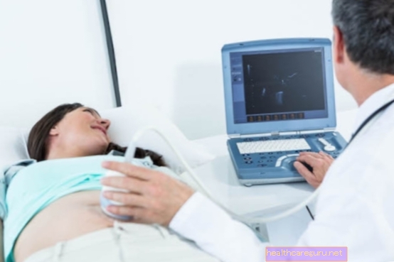
Thus, to identify the septate uterus, the gynecologist can indicate the performance of imaging exams such as ultrasound, endocervical curettage and hysterosalpingography.
Often the septate uterus is confused with the bicornuate uterus, which is when the uterus is not fully connected to the cervix, and the differentiation between these two changes can be made through 3D ultrasound or an exam called hysteroscopy. See more about the bicornuate uterus.
Is it possible to get pregnant with a septate uterus?
Pregnancy with a septate uterus is, in most cases, difficult, because as the uterus is divided, there are not enough blood vessels to allow the embryo to be implanted in the uterus, and there is no pregnancy.
In the case of implantation, the presence of the septum can interfere with the supply of nutrients and oxygen to the fetus, which can directly interfere with its development and favor the occurrence of spontaneous abortions. In addition, as the space is smaller due to the presence of the septum, the baby's growth can also be hampered.
How the treatment is done
Treatment for septate uterus must be guided by a gynecologist and is usually done through surgery that removes the wall that divides the uterus into two parts. This removal is done through a surgery called surgical hysteroscopy, where a device is inserted through the vagina into the uterus to remove the septum.
This procedure is done with general or spinal anesthesia, lasts about 30 minutes to 1 hour, and the woman can go home on the day of the surgery. However, it is normal for vaginal bleeding to occur for up to 6 weeks after surgery, and it is usually necessary to take medications to relieve pain and decrease inflammation in the uterus, in addition to antibiotics to prevent infections.
The precautions that must be taken in the 2 weeks following the surgery are to avoid making physical efforts, such as picking up heavy objects or working out, not having intimate contact and avoid taking a bath in the pool and the sea. In case of fever, pain, heavy vaginal bleeding or a bad-smelling discharge, you should see your doctor.
In general, about 8 weeks after surgery the woman is reevaluated to check the result of the surgery and be released to become pregnant. Check out more details about surgical hysteroscopy.
Created by: Tua Saúde Editorial Team
Bibliography>
- FERREIRA, Adilson C .; FILHO, Francisco M .; NICOLAU, Luis G .; GALLARRETA, Francismo M. P. Three-dimensional ultrasonography in gynecology: uterine malformations. Radiol Bras. Vol 40. 2nd ed; 131-136, 2007
- BRAZILIAN FEDERATION OF GYNECOLOGY AND OBSTETRICS ASSOCIATIONS. Uterine Factor Infertility. 2013. Available at:. Accessed on 02 Sep 2020