Content
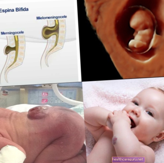
Myelomeningocele is the most serious type of spina bifida, in which the baby's spine bones do not develop properly during pregnancy, causing the appearance of a pouch on the back containing the spinal cord, nerves and cerebrospinal fluid.
Generally, the appearance of the myelomeningocele pouch is more frequent at the bottom of the back, but it can appear anywhere on the spine, causing the child to lose the sensitivity and function of the limbs below the location of the alteration.
Myelomeningocele has no cure because, although it is possible to reduce the bag with surgery, the lesions caused by the problem cannot be completely reversed.
Main symptoms
The main symptom of myelomeningocele is the appearance of a pouch on the baby's back, however, other signs include:
- Difficulty or absence of movement in the legs;
- Muscle weakness;
- Loss of sensitivity to heat or cold;
- Urinary and fecal incontinence;
- Malformations in the legs or feet.
Usually, the diagnosis of myelomeningocele is made right at birth with the observation of the bag on the baby's back. In addition, the doctor usually requests neurological exams to check for any nerve involvement.
What causes myelomeningocele
The cause of myelomeningocele is not yet well established, however it is believed that it is the result of genetic and environmental factors, and is usually related to a history of spinal malformations in the family or folic acid deficiency.
In addition, women who used certain anticonvulsant medications during pregnancy, or have diabetes, for example, are more likely to have myelomeningocele.
To prevent myelomeningocele, it is important for pregnant women to supplement folic acid before and during pregnancy, as in addition to avoiding myelomeningocele, it prevents premature delivery and pre-eclampsia, for example. See how folic acid supplementation should be done during pregnancy.
How the treatment is done
Treatment of myelomeningocele is usually started within the first 48 hours after birth with surgery to correct the alteration in the spine and prevent the appearance of infections or new lesions in the spinal cord, limiting the type of sequelae.
Although treatment for myelomeningocele with surgery is effective in curing the baby's spine injury, it is not able to treat the sequelae that the baby has had since birth. That is, if the baby was born with paralysis or incontinence, for example, it will not be cured, but it will prevent the appearance of new sequelae that could arise from spinal cord exposure.
How is the surgery done
Surgery to treat myelomeningocele is usually done in the hospital under general anesthesia and should ideally be done by a team that contains a neurosurgeon and a plastic surgeon. That's because it usually follows the following step-by-step:
- The neurosurgeon closes the spinal cord;
- The back muscles are closed by a plastic surgeon and the neurosurgeon;
- The skin is closed by the plastic surgeon.
Often, as there is little skin available at the site of the myelomeningocele, the surgeon needs to remove a piece of skin from another part of the baby's back or bottom, to perform an excerpt and close the opening in the back.
In addition, most babies with myelomeningocele can also develop hydrocephalus, which is a problem that causes excessive accumulation of fluid inside the skull and, therefore, it may be necessary to have a new surgery after the first year of life to place a system that helps to drain fluids to other parts of the body. Learn more about how hydrocephalus is treated.
Is it possible to have surgery on the uterus?
Although it is less frequent, in some hospitals, there is also the option of having surgery to end myelomeningocele before the end of pregnancy, still inside the pregnant woman's uterus.
This surgery can be done at around 24 weeks, but it is a very delicate procedure that should only be done by a well-trained surgeon, which ends up making the surgery more expensive. However, the results of surgery in the uterus appear to be better, as there is less chance of new spinal cord injuries during pregnancy.
Physiotherapy for myelomeningocele
Physiotherapy for myelomeningocele must be done during the baby's growth and development process to maintain the amplitude of the joints and avoid muscle atrophy.
In addition, physiotherapy is also a great way to encourage children to deal with their limitations, as in the case of paralysis, allowing them to have an independent life, using crutches or a wheelchair, for example.
When you go back to the doctor
After the baby is discharged from the hospital it is important to go to the doctor when symptoms such as:
- Fever above 38ºC;
- Lack of desire to play and apathy;
- Redness at the surgery site;
- Decreased strength in unaffected limbs;
- Frequent vomiting;
- Dilated soft spot.
These symptoms can indicate serious complications, such as infection or hydrocephalus, so it is important to go to the emergency room as soon as possible.

.jpg)